
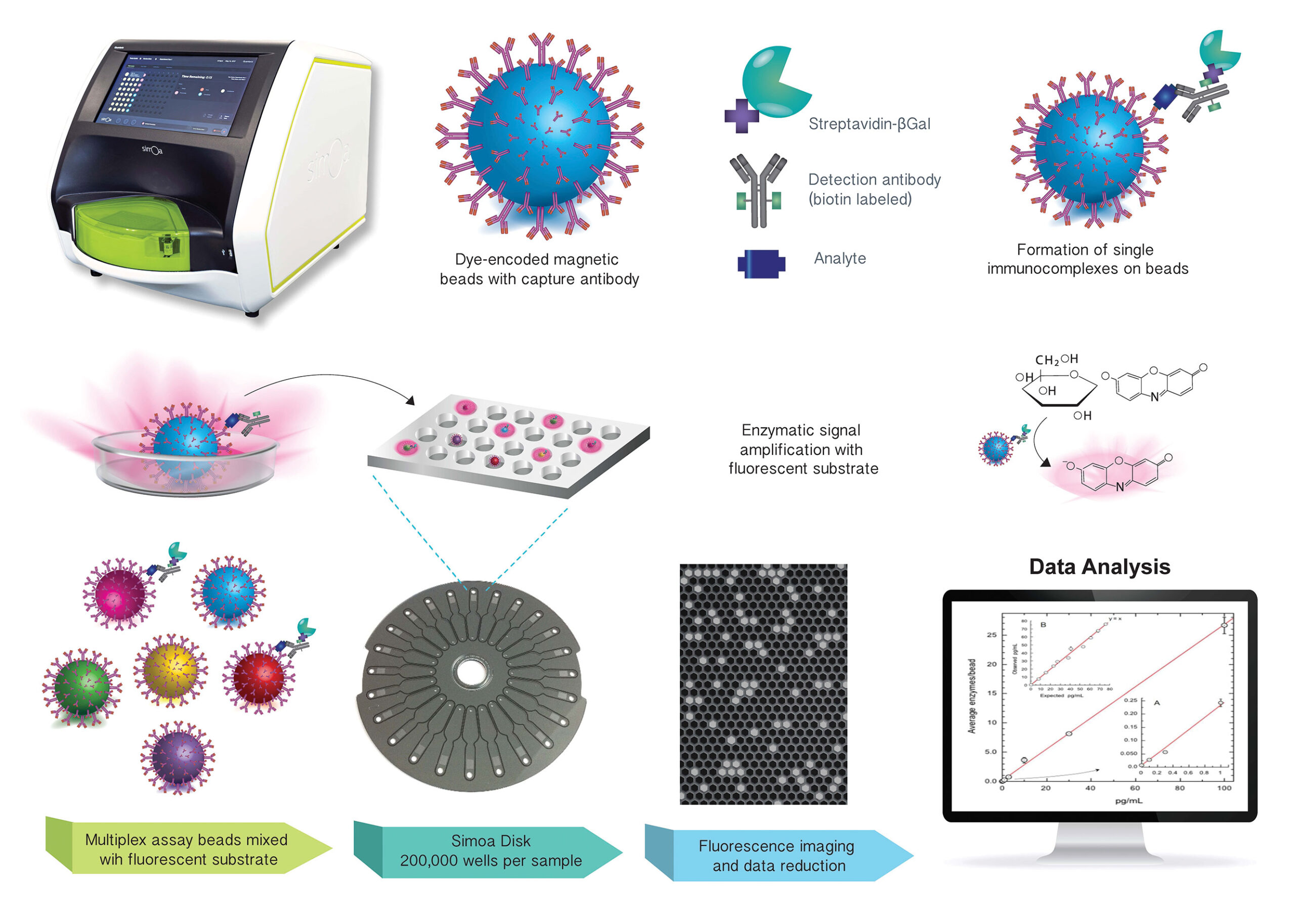
Research into Alzheimer’s disease increasingly depends on reliable methods for measuring biological markers associated with neurological changes. These biomarkers help scientists investigate amyloid accumulation, tau pathology, and neuronal injury, providing a more detailed understanding of disease-related processes. As research expands, laboratories need analytical methods that can measure specific proteins accurately and consistently across different sample types.
Selecting an appropriate alzheimer biomarker assay requires evaluating several factors, including analytical sensitivity, specificity, precision, sample compatibility, and workflow requirements. Different technologies offer distinct detection principles and performance characteristics. Understanding these differences helps researchers choose suitable methods for studying Alzheimer’s-related biomarkers and generating reproducible laboratory data.
What Are Alzheimer’s Disease Biomarkers?
Biomarkers are measurable biological characteristics that provide information about normal physiological processes, disease-related changes, or responses to interventions.
In Alzheimer’s research, biomarkers are used to investigate several processes associated with the condition, including:
- Amyloid-beta accumulation.
- Abnormal tau phosphorylation and aggregation.
- Neuronal and axonal injury.
- Changes in neurological function.
Biomarkers can be measured in different biological samples, including cerebrospinal fluid (CSF), plasma, and serum. Some can also be evaluated through imaging techniques.
Each biomarker provides different information. For this reason, researchers often combine multiple measurements rather than relying on a single result.
Major Biomarkers Used in Alzheimer’s Research
Understanding the biological target is the first step in selecting an appropriate analytical method.
Amyloid-Beta 42 and Amyloid-Beta 40
Amyloid-beta peptides are produced through the processing of amyloid precursor protein.
Amyloid-beta 42 is particularly associated with amyloid plaque formation in the brain. Amyloid-beta 40 is also commonly measured, and the ratio of amyloid-beta 42 to amyloid-beta 40 can provide additional information about amyloid-related changes.
Researchers investigate these markers to understand amyloid pathology and its relationship with other Alzheimer’s-associated processes.
Phosphorylated Tau 181
Phosphorylated tau 181, commonly called p-tau 181, is a modified form of tau protein phosphorylated at the threonine 181 position.
It is studied because its levels in CSF and blood may reflect biological changes associated with Alzheimer’s disease.
Laboratories measuring p-tau 181 need assays with appropriate antibody specificity to distinguish the intended phosphorylated form from other tau variants.
Phosphorylated Tau 217
P-tau 217 is another phosphorylated tau marker that has attracted considerable research interest.
Studies have investigated its relationship with Alzheimer’s pathology and its potential value in blood-based biomarker strategies.
Total Tau
Total tau refers to measurements of tau protein without restricting detection to a specific phosphorylation site.
It is generally investigated as a marker associated with neuronal injury. Its interpretation differs from that of phosphorylated tau measurements.
Neurofilament Light Chain
Neurofilament light chain (NfL) is a structural protein associated with neurons, particularly their axons.
Increased NfL concentrations can reflect axonal injury and are studied across several neurological conditions. It is not specific to Alzheimer’s disease.
Why Analytical Methods Matter in Biomarker Research
A biomarker can only provide useful research information when the analytical method measures it reliably.
Different assays may produce different results because of variations in antibody design, calibration, detection chemistry, sample preparation, and measurement range.
A suitable analytical method should demonstrate:
- Appropriate sensitivity for the target concentration.
- Specificity for the intended biomarker.
- Consistent performance across repeated measurements.
- Compatibility with the required biological sample.
- Reliable calibration and quality-control procedures.
- A measurement range suitable for the study population.
These characteristics are important when comparing research findings, conducting longitudinal studies, or combining data from different laboratories.
Common Analytical Approaches for Alzheimer’s Biomarker Testing
Several technologies are used to measure proteins associated with Alzheimer’s disease. Each has its own advantages, limitations, and practical requirements.
1. Enzyme-Linked Immunosorbent Assay (ELISA)
ELISA is a widely used immunoassay technique for measuring proteins and other biological molecules.
It relies on antibodies that capture and recognize the target analyte. An enzyme-based reaction then produces a measurable signal.
How it works:
- A capture antibody binds to the target protein.
- The sample is incubated under defined assay conditions.
- A detection antibody recognizes another part of the target.
- An enzyme-based reaction generates a signal.
- The signal is measured and compared with a calibration curve.
Advantages:
- Established laboratory methodology.
- Suitable for many protein research applications.
- Available in different assay formats.
- Can support quantitative measurements.
Considerations:
- Some formats involve multiple manual steps.
- Incubation and washing conditions must be controlled.
- Analytical sensitivity varies between kits.
- Batch processing may influence laboratory workflow.
ELISA is often suitable for research applications where the laboratory can maintain standardized procedures and appropriate quality controls.
2. Chemiluminescence Immunoassay (CLIA)
CLIA combines antibody-based recognition with a chemical reaction that produces light.
The intensity of the emitted light is measured by a compatible analyzer and interpreted using a calibration system.
How it works:
- Antibodies recognize the target biomarker.
- The target and relevant antibodies form an immune complex.
- A chemiluminescent reaction generates light.
- The analyzer detects the signal.
- Software or an established calculation procedure determines the analyte concentration.
Advantages:
- Can support sensitive protein detection.
- Suitable for automated laboratory workflows.
- Allows quantitative measurement within a validated range.
- Can reduce some forms of manual handling.
Considerations:
- Performance depends on the specific reagent system.
- Instrument and assay compatibility must be confirmed.
- Calibration and quality-control procedures are essential.
- Different platforms may produce non-interchangeable results.
CLIA can be useful when laboratories need automated measurement of Alzheimer’s-related proteins, provided the selected assay has been validated for the intended application.
3. Electrochemiluminescence Immunoassay (ECLIA)
Electrochemiluminescence immunoassay uses an electrically initiated reaction to produce light.
The generated signal is measured to estimate the concentration of the target biomarker.
Advantages:
- Can support sensitive protein measurement.
- Offers an established approach for automated testing.
- Can be applied to different immunoassay formats.
Considerations:
- Requires compatible detection systems and reagents.
- Performance depends on assay design and validation.
- Results may differ from those generated by other analytical platforms.
ECLIA is used in various biomarker research and laboratory testing applications.
4. Mass Spectrometry
Mass spectrometry is an analytical technique that identifies and quantifies molecules according to their mass-to-charge characteristics.
In protein biomarker research, it can be used to investigate specific peptides and protein-related molecular features.
Advantages:
- Provides molecular information.
- Can support highly specific analytical investigations.
- Useful for research involving multiple molecular targets.
Considerations:
- Requires specialized instrumentation and expertise.
- Sample preparation can be complex.
- Method development and data analysis may require substantial resources.
- It may not be practical for every routine laboratory workflow.
Mass spectrometry can complement immunoassay methods, particularly when researchers need additional molecular characterization.
5. Fluorescence-Based Immunoassays
Fluorescence immunoassays use fluorescent labels to generate measurable signals when antibodies bind to their targets.
These methods can be adapted for different research applications and assay formats.
Advantages:
- Flexible detection approaches.
- Suitable for various biomolecular targets.
- Can be adapted for different laboratory workflows.
Considerations:
- Fluorescence background can affect measurement.
- Signal stability and reagent properties require attention.
- Sensitivity varies according to assay design and instrumentation.
Comparing Analytical Approaches
The following table summarizes the general characteristics of common methods used in Alzheimer’s biomarker research.
| Analytical method | Detection principle | General strengths | Important considerations |
|---|---|---|---|
| ELISA | Enzyme-generated signal | Established and widely used | Manual steps and assay-dependent sensitivity |
| CLIA | Chemically generated light | Automation and quantitative detection | Requires compatible reagents and analyzer |
| ECLIA | Electrically initiated light emission | Sensitive immunoassay detection | Platform-specific requirements |
| Mass spectrometry | Mass-to-charge measurement | Molecular characterization | Specialized equipment and expertise |
| Fluorescence immunoassay | Fluorescent signal | Flexible assay formats | Background signal and label stability |
There is no universally best method for every Alzheimer’s biomarker. The appropriate choice depends on the target, sample type, research objective, laboratory resources, and validated analytical performance.
Key Performance Characteristics of an Alzheimer Biomarker Assay
Selecting an assay involves more than identifying its detection technology. Laboratories should review the complete analytical performance profile.
Analytical Sensitivity
Sensitivity describes the ability of an assay to detect low concentrations of the target.
Important parameters include:
- Limit of blank (LoB): The highest apparent concentration expected in samples without the target.
- Limit of detection (LoD): The lowest concentration that can be distinguished from the blank with defined confidence.
- Limit of quantification (LoQ): The lowest concentration that can be measured with acceptable precision and accuracy.
- Analytical measurement range: The concentration interval over which the assay provides reliable quantitative results.
Sensitivity is particularly important for blood-based Alzheimer’s biomarkers, where some target proteins may be present at low concentrations.
Analytical Specificity
Specificity describes how effectively a method measures the intended target without substantial interference from related substances.
For phosphorylated tau assays, antibody specificity is especially important.
A p-tau 181 method should recognize the phosphorylation site at threonine 181 and be evaluated for cross-reactivity with other tau forms.
Precision and Reproducibility
Precision evaluates the consistency of repeated measurements.
Laboratories should examine:
- Within-run precision.
- Between-run precision.
- Between-day variation.
- Reproducibility across operators or laboratories, when relevant.
These characteristics are essential for studies involving repeated measurements or comparisons between research sites.
Accuracy and Bias
Accuracy concerns how closely a measurement reflects an appropriate reference value.
Bias describes systematic differences between the measured result and the reference.
Researchers should consider the availability of suitable reference materials and method-comparison evidence when evaluating assay accuracy.
Interference and Matrix Effects
Biological samples contain many substances that may affect analytical performance.
Potential interferents include hemoglobin, lipids, bilirubin, heterophilic antibodies, and other matrix components.
Interference studies help determine whether these substances affect the assay under expected testing conditions.
Sample Types Used in Alzheimer’s Biomarker Research
The sample type can influence assay selection and analytical performance.
Cerebrospinal Fluid
CSF surrounds the brain and spinal cord and contains proteins that can reflect neurological processes.
It is commonly used in Alzheimer’s biomarker research, particularly for amyloid-beta and tau measurements.
CSF collection generally requires lumbar puncture, which is more invasive than blood collection.
Plasma
Plasma is obtained from anticoagulated blood and is increasingly investigated for Alzheimer’s-related biomarkers.
Blood-based testing may make large-scale research and repeated sample collection more practical.
However, plasma has a complex composition, and the concentration of certain biomarkers can be low. Assays must therefore demonstrate suitable performance in this specific matrix.
Serum
Serum is collected after blood has clotted.
Its composition differs from plasma, so analytical results and assay performance may vary between these sample types.
Researchers should verify sample compatibility rather than assuming that a method validated for plasma will also work with serum.
The Role of Automation in Alzheimer’s Biomarker Testing
Automation can improve consistency by standardizing several laboratory steps.
Automated immunoassay analyzers may manage reagent dispensing, incubation, washing, signal detection, and result calculation.
In CLIA workflows, automation can help reduce some sources of manual variation and support higher testing throughput.
When evaluating a platform such as the Shine i2910 Chemiluminescence Immunoassay Analyzer, laboratories should confirm that the intended Alzheimer’s biomarker assay is compatible with the instrument and validated for the required sample type.
Important considerations include:
- Reagent and assay compatibility.
- Supported sample types.
- Analytical performance of the complete assay system.
- Testing capacity and workflow requirements.
- Calibration and quality-control procedures.
- Maintenance and technical support.
An analyzer’s general chemiluminescence capabilities do not independently establish its suitability for every Alzheimer’s biomarker. The instrument and reagent system must be assessed together.
Quality Control and Standardization
Reliable biomarker measurements require ongoing quality monitoring.
Calibration
Calibration establishes the relationship between an assay signal and the concentration of the target analyte.
Laboratories should follow the assay’s calibration instructions and use appropriate materials covering the intended measurement range.
Internal Quality Control
Internal quality-control materials help laboratories monitor assay performance during routine testing.
Controls at different concentration levels can help identify shifts, trends, or unexpected variation.
External Quality Assessment
External quality assessment allows laboratories to compare results with other participating facilities.
Where suitable programs are available, they can help identify differences between methods and support improvements in analytical practice.
Standardized Procedures
Consistent procedures for sample collection, processing, storage, and analysis are essential.
Laboratories should document reagent batches, calibration results, control measurements, instrument maintenance, and corrective actions.
Challenges in Comparing Alzheimer’s Biomarker Assays
Comparing analytical methods can be difficult because different platforms may use different reagents, calibration systems, and detection principles.
Differences in Antibody Design
Antibody pairs may recognize different regions or forms of a protein, leading to differences in measured concentrations.
Calibration Differences
Different calibrators and assigned values can affect how results are reported.
Sample Matrix Effects
An assay may perform differently in CSF, plasma, and serum.
Variations in Measurement Range
Methods may have different lower and upper quantification limits, making direct comparisons difficult.
Lack of Interchangeability
Results from separate platforms should not automatically be treated as equivalent. Appropriate method-comparison studies and validation are necessary before combining measurements.
How to Select the Right Analytical Method
A practical selection process begins with the research objective and works backward to the analytical requirements.
Define the target biomarker. Determine whether the study focuses on amyloid-beta, phosphorylated tau, total tau, NfL, or multiple targets.
Choose the sample type. Confirm whether CSF, plasma, or serum will be used.
Set analytical requirements. Establish the expected concentration range, required sensitivity, and acceptable precision.
Compare detection technologies. Evaluate ELISA, CLIA, ECLIA, mass spectrometry, and fluorescence-based approaches according to the study’s needs.
Review validation evidence. Examine specificity, accuracy, precision, interference, and sample stability data.
Assess laboratory workflow. Consider equipment availability, throughput, reagent handling, training, and maintenance.
Establish quality procedures. Define calibration, internal controls, documentation, and result-review requirements.
This structured approach helps laboratories select methods based on evidence rather than relying on a single specification.
Future Trends in Alzheimer’s Biomarker Assay Development
Research into Alzheimer’s disease biomarkers continues to evolve.
More Sensitive Detection Technologies
Advances in antibody engineering, signal generation, and analytical instrumentation may improve the measurement of low-abundance proteins.
Expanded Blood-Based Biomarker Research
Blood-based assays are being investigated as more accessible options for biomarker studies and potentially broader clinical applications.
Multiplex Testing
Multiplex approaches aim to measure several biomarkers within a coordinated analytical workflow.
Combining amyloid, tau, and neuronal injury markers may provide a broader understanding of disease-related biology.
Greater Standardization
Improved reference materials, calibration systems, and external quality-assessment approaches may help make results more comparable between laboratories.
Integration With Other Research Data
Biomarker measurements may increasingly be evaluated alongside imaging, genetics, and clinical information to investigate complex disease processes.
These developments will require continued analytical and clinical validation before new methods can be adopted for specific applications.
Conclusion
Alzheimer’s biomarker research uses several analytical approaches to investigate amyloid accumulation, tau pathology, and neuronal injury. ELISA, CLIA, ECLIA, mass spectrometry, and fluorescence-based immunoassays each offer distinct detection principles and practical considerations.
Choosing an appropriate assay requires evaluating analytical sensitivity, specificity, precision, accuracy, sample compatibility, calibration, and workflow requirements. Automated CLIA platforms may support consistent testing when paired with compatible, validated reagent systems.
By selecting methods that match the research objective and maintaining rigorous quality-control procedures, laboratories can generate more reproducible data and contribute to a clearer understanding of Alzheimer’s disease and other neurodegenerative conditions.